


Scanner de ultrassom doppler portátil
$2100-3500 /Set/Sets
| Tipo de pagamento: | T/T |
| Incoterm: | FOB |
| Quantidade de pedido mínimo: | 5 Set/Sets |
| transporte: | Ocean,Land,Air,Express |
| porta: | Shenzhen,Ningbo,Shanghai |

$2100-3500 /Set/Sets
| Tipo de pagamento: | T/T |
| Incoterm: | FOB |
| Quantidade de pedido mínimo: | 5 Set/Sets |
| transporte: | Ocean,Land,Air,Express |
| porta: | Shenzhen,Ningbo,Shanghai |
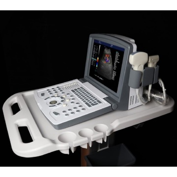
Modelo: MDK-680
marca: Ultra -som unido
Lugar De Origem: China
Tipo De Poder: Eletricidade
Serviço De Garantia: 1 ano
Serviço Pós-venda: Peças de reposição grátis, Suporte técnico online
Material: metal, plástico
Classificação De Dispositivos Médicos: Classe II
Color: white
Brand: United Ultrasound
Monitor Size: 12 inch
Gray Scale: 256 levels
Depth: ≥300mm
Channels: 32
Operating System: Windows 7
| Unidades de venda | : | Set/Sets |
| Tipo de pacote | : | Carton |
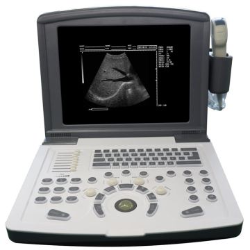
Scanner de ultrassom doppler portátil para colorido para vascular (mdk-680)
A ultrassonografia vascular do Doppler de cor é um método de exame amplamente usado em nossa vida diária. Esse método de exame pode verificar principalmente se há trombose no corpo do paciente ou se há doença na artéria do paciente. Também pode verificar se a artéria do paciente está bloqueada.
O ultrassom de Doppler colorido pode detectar se há placa na artéria e se a placa causa estenose ou oclusão. O ultrassom vascular do Doppler de cor é amplamente utilizado na prática clínica, usada principalmente para detectar as seguintes doenças: primeiro, se há uma doença do sistema venoso. Se há trombose na veia, se há condição de válvula, se há insuficiência. Segundo, verifique as artérias quanto a doenças. Como arteriosclerose, trombose, estenose e assim por diante.
Nossos produtos:
Scanner de ultrassom preto e branco
Scanner de ultrassom de Doppler colorido
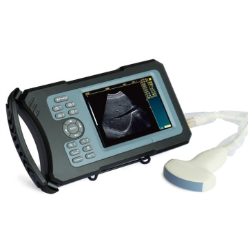
Scanner de ultrassom veterinário


|
No. |
Item |
Index |
|
<1> |
Depth |
≥300mm |
|
<2> |
Lateral Resolution |
≤1mm (Depth≤80mm) ≤2mm (80< Depth≤130mm) |
|
<3> |
Axial Resolution |
≤1mm (Depth≤80mm) ≤2mm (80< Depth≤130mm) |
|
<4> |
Blind Area |
≤4 mm |
|
<5> |
Geometry Position Precision |
horizontal≤5% vertical≤5% |
|
<6> |
Language |
English/Chinese |
|
<7> |
Channels |
32 |
|
<8> |
Displayer |
12” LED |
|
<9> |
External Display |
PAL, VGA, USB |
|
<10> |
Gray Scale |
256 levels |
|
<11> |
Voltage |
AC220V ±10% |
|
<12> |
Operating System |
Windows 7 |
|
<13> |
Scanning Mode |
B, B/B, 4B, B/M, M, B+C, B+D, B+C+D, PDI, CF, PW |
|
<14> |
Probe |
Probe sockets: 2 Probe frequency: 2.0MHz ~ 10.0MHz, 8-step frequency conversion |
|
<15> |
Adjustment parameters of color blood flow image |
Doppler frequency, sampling frame position and size, baseline, color gain, deflection angle, wall filtering, cumulative times, etc |
|
<16> |
Signal processing
|
With dynamic filtering and quadrature demodulation With total gain adjustment Gain adjustment: 8-segment TGC The total gain of Type B, Type C and Type D can be adjusted respectively B/W image gain and color blood flow gain are adjustable respectively |
|
<17> |
Doppler |
Doppler baseline adjustment level 6 Pulse repetition frequency can be adjusted separately: CFM PWD With D linear speed regulation |
|
<18> |
Digital beam forming |
Continuous dynamic focusing of digital beam forming image Full range dynamic aperture of image Dynamic tracing of the whole image Weighted Sum of Image Whole Process Receiving Delay Support half step scanning and ± 10 ° linear receiving deflection angle Multi beam parallel processing technology |
|
<19> |
Basic measurement and calculation function |
Basic measurement in mode B: distance, angle, perimeter and area, volume, stenosis rate, histogram, cross-section |
|
Basic measurement of M-mode: heart rate, time, distance and speed |
||
|
Doppler measurement: time, heart rate, speed, acceleration |
||
|
<20> |
Gynecological measurement and calculation function |
Measurement and calculation of uterus, left ovary, right ovary, left follicle, right follicle, etc |
|
<21> |
Obstetric measurement and calculation function |
G.A, EDD, BPD-FW, FL, AC, HC, CRL, AD, GS, LMP,HL,LV,OFD |
|
<22> |
Urology measurement and calculation function
|
Measurement and calculation of left kidney, right kidney, bladder, residual urine volume, prostate, prostate specific antigen predicted value PPSA, prostate specific antigen density PSAD, etc |
|
<23> |
Product Size |
289×304×222mm |
|
<24> |
Carton Size |
395×300×410mm |
|
<25> |
N.W./ G.W. |
6kg/ 7kg |


Privacy statement: Your privacy is very important to Us. Our company promises not to disclose your personal information to any external company with out your explicit permission.
Fill in more information so that we can get in touch with you faster
Privacy statement: Your privacy is very important to Us. Our company promises not to disclose your personal information to any external company with out your explicit permission.


